
Osteoporosis screening by dental panoramic X-ray imaging
Osteoporosis and
bone fractures
Osteoporosis is a disease characterized by bone brittleness that can lead to fragility fractures (Reference 1).
The onset of osteoporosis is closely related to age and gender, with the incidence rate increasing markedly in women in their 50s and beyond. Accordingly, in Japan, which is facing general population aging, the number of patients with osteoporosis is estimated to be 13 to 15 million or more (Reference 2).
Bone fractures due to osteoporosis often occur in the proximal femur, vertebral bodies (lumbar vertebrae), and distal radius (wrist). Proximal femur fractures, in particular, are reportedly associated with mortality rates of up to 10% one year after the fracture (Reference 3). Not only proximal femoral fractures, but also vertebral body fractures lead to markedly decreased quality of life (QOL) and increased risks of death.
In addition, there have been an increasing number of cases where the patient with fragility fractures due to osteoporosis is unable to restore their social life even after surgery and eventually becomes bedridden and requires long-term care. Osteoporosis is estimated to result in 2 trillion yen of medical and nursing care costs in 2025. These costs are expected to continue to rise with increasing population aging (References 4 and 5).
Osteoporosis
screening by dental
panoramic X-ray imaging
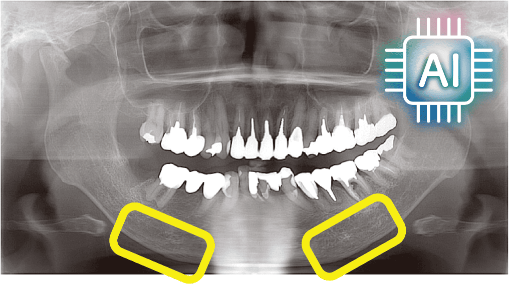
In dentistry, there has been increasing attention paid to a screening method for potential patients with osteoporosis to be used by the dentist observing the morphology of the cortical bone at the lower margin of the mandible using panoramic X-ray images taken for dental treatment (Reference 6).
However, this method is not widely used in dental clinics because of the lack of dedicated equipment and software. We at MEDIA Co., Ltd. (https://www.media-inc.co.jp [in Japanese]) have developed software that assists the dentist in determining the likelihood of osteoporosis based on an assessment of jawbone fragility by automatically analyzing dental panoramic X-ray images with artificial intelligence (AI) image processing technology.
This software is the first AI-programmed medical device approved for dentistry in Japan (References 7 and 8). We also run a website for healthcare professionals that supports medical-dental collaboration regarding osteoporosis and drug-related osteonecrosis of the jaw caused by antiresorptive drugs.
AI software for
osteoporosis
screening in dentistry
This software assists the dentist in osteoporosis screening by inputting digital panoramic X-ray images and measuring/analyzing the following parameters to evaluate “fragility changes” of the jawbone:
1) Detects the cortical bone at the lower edge of the mandible and indicates a measurement point near the mental foramen.
2) Measures the mandibular cortical width (MCW), i.e., thickness of the cortical bone.
3) Calculates the mandibular cortex morphology index (MCMI), an indicator of the morphology (degree of osteoporosis) of the cortical bone.
4) Determines the mandibular cortical index (MCI) to classify the mandibular cortical bone morphology.
〈references〉
1) Assessment of fracture risk and its application to screening for postmenopausal osteoporosis: report of a WHO study group [meeting held in Rome from 22 to 25 June 1992]. WHO technical report series 1994,843.
2) Noriko Yoshimura, et al. Trends in osteoporosis prevalence over a 10-year period in Japan: the ROAD study 2005-2015. J Bone Miner Metab. 40(5):829-838, 2022.
3) Sakamoto K, Nakamura T, Hagino H, Endo N, Mori S, Muto Y, Harada A, Nakano T, Yamamoto S, Kushida K, Tomita K, Yoshimura M, Yamamoto H. Report on the Japanese Orthopaedic Association's 3-year project observing hip fractures at fixed-point hospitals. J Orthop Sci. 2006Mar;11(2):127-34.
4) Hagino H, Kondo A, Oeki M. Medical costs for the treatment of osteoporotic fractures. THE BONE . 2009;23(2):47-51.
5) Harada A, Matsui Y, Takemura M, Ito Z, Wakao N, Ota T (2005) Cost-utility analysis of osteoporosis. Nippon Ronen Igakkai Zasshi. Jpn J Geriatr42:596-608
6) Taguchi A, Tanaka R, Kakimoto N, et al. Clinical guidelines for the application of panoramic radiographs in screening for osteoporosis. Oral Radiology. 2021;37(2):189-208.
7) Maruo N, Toyoshima K, Katsumata A. Kagawa prefecture’s medical dental collaboration framework to prevent osteoporosis using panoramic radiography (in Japanese). Japanese Journal of Dental Practice Administration. 2019;53(4):206-211.
8) Katsumata A. AI-based osteoporosis screening using dental panoramic radiography (in Japanese). Journal of Japan Osteoporosis Society. 2023;9:461-466.
Akitoshi Katsumata

1987: Graduated from the Department of Dentistry, School of Dentistry, Asahi University.
2011: Appointed professor at Asahi University (current position).
Japan Dental Artificial Intelligence Study Group (Representative Director)
Japanese Society of Dental Radiology and more.
After graduating from the Asahi University School of Dentistry, he served as an assistant, lecturer, and associate professor for the Dental Radiology Course and was then appointed as a professor. He is currently working on clinical practice for diagnostic imaging and research and development of artificial intelligence software.